| Session: | SE63-SE78-Sports Medicine/Arthroscopy Scientific Exhibits |
| Date/Time: | Wednesday, Mar 20, 2013, 7:00 AM – 6:00 PM Scientific Exhibits occur Tuesday through Saturday. Click here to view all Scientific Exhibits. |
| Location | McCormick Place, Academy Hall B |
| Presentation Number: | SE73 |
| Title: | The Anterolateral Ligament of the Knee: Anatomy, Radiology, Biomechanics and Clinical Implications |
| Classification: | +Knee- Ligaments (excluding ACL) (Sports Med/Arthro) |
| Keywords: | ACL Deficient; Knee Arthroscopy; MRI / Imaging; Anatomy / Biomechanics |
| Author(s): | Steven A. Claes, MD, Pellenberg, Belgium Stijn Bartholomeeusen, MD, Malle, Belgium Evie E. Vereecke, PhD, Kortrijk, Belgium Jan M. Victor, MD, GENT, Belgium Peter Verdonk, MD, PhD, Ghent, Belgium Johan Bellemans, MD, Langdorp, Belgium |
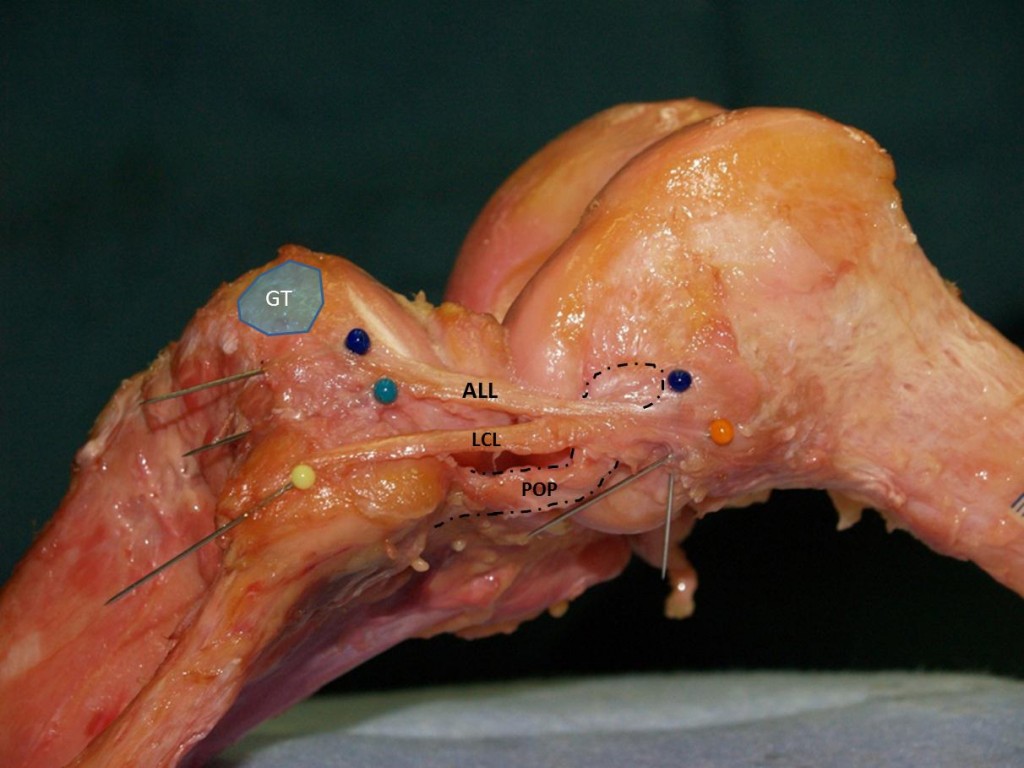
| Abstract: | Introduction: In 1879 dr. Segond described the existence of a “pearly, fibrous band” at the anterolateral aspect of the human knee attached to the eponymous fracture. To date, the enigma surrounding this anatomical structure is reflected in confusing names like “(mid-third) lateral capsular ligament”, “capsulo-osseous layer of the ITB”, or “anterolateral ligament”, and no clear function has yet been attributed to it. Therefore, the goal of this study was to provide a precise anatomic description (1) of this anterolateral ligament (ALL), describe its biomechanics (2) in computer-navigated sequential cutting experiments, characterize the ALL on knee MRIs (3) and delineate the Segond fracture (4) as a bony avulsion of the ALL. Methods: (1) The ALL was investigated in 35 human cadaveric knees; its dimensions and relation with anatomical landmarks were recorded. (2) Navigated knee kinematics were obtained from 10 fresh-frozen cadavers in the native knee, after sequential cutting of the two bundles of the ACL and the ALL respectively. (3) The aspect of the ALL was studied on MRI images of 350 ACL injured subjects. (4) Segond fracture characteristics were studied on MR images of 26 subjects and compared with the anatomical findings. Results: (1) The ALL was found in all knees; it invariably originated on the lateral femoral epicondyle, in close relation to the lateral collateral ligament. The ALL showed an oblique course to the tibia (posterior and proximal to Gerdy’s tubercle), firmly attached to the lateral meniscus. (see figure) (2) The ALL was found to be an important internal rotatory stabilizer of the knee, especially in flexion angles between 30° and 60° and this in both the ACL-intact and -deficient knee. With regard to the pivot-shift, cutting of the ALL increased the pivot-shift with a least 1 grade, regardless of the condition the ACL. All knees with both ALL and ACL cut, resulted in a gross, grade III pivot-shift, while removal of both ACL bundles alone did never, the latter only delivering a grade I pivot-shift in 40% of the knees. (3) The ALL was found on MRI in 95,3% of the ACL-injured subjects. Lesions of the ALL were noted in 79,2% of these patients, mostly distal. (4) The Segond fracture was delineated as a bony avulsion of the ALL. Discussion and Conclusion: Previously undescribed, the ALL was found to be a distinct anatomical structure of the human knee with definite biomechanical properties. Although the classic concept of anterolateral rotatory knee instability developed by Hughston, already implied a combination of injuries to both the ACL and the “anterolateral stabilizing structures”, this interesting notion has seemingly become obsolete under the boom of arthroscopic surgery . However, the high incidence of ALL lesions on MR images of ACL-injured subjects and its causative relationship with a high-grade pivot-shift, yield exciting new insights related to the interaction between ALL and ACL, and stimulate further research in this common field of knee instability. For illustration, a multi-media presentation of Segond refixation and ALL reconstruction procedures will be displayed. In conlusion, the ALL is a distinct anatomical structure with definite biomechanical properties. Its recognition yields new insights for the diagnosis and treatment of common knee instability patterns, previously attributed to isolated injuries of the ACL or one of its bundles.  |