Abstract
INTRODUCTION:
The sternoclavicular joint is of clinical importance. However, there is scant information in the literature regarding one ligament of this area, the costoclavicular ligament (CCL).
MATERIAL AND METHODS:
In order to further elucidate this structure, 10 adult formalin-fixed cadavers (17 sides) underwent dissection of the CCL. Once the CCL was identified, measurements were made of its dimensions and observations made of its anatomy. Next, ranges of motion were performed of the upper extremity and the CCL observed for tension or laxity.
RESULTS:
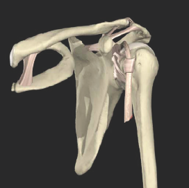
Of the 17 sternoclavicular regions examined 16 (94%) were found to possess a CCL. The average medial and lateral lengths, width and thickness were 1, 2, 1.2, 0.340 cm, respectively. The width of the CCL was statistically smaller in women that in men. The majority of ligaments were single structures traveling from the inferior surface of the medial clavicle just lateral and sometimes-fused (12.5%) to the lateral edge of the sternoclavicular joint. These fibers then terminated on the medial end of the first rib and first costal cartilage (75%) or exclusively onto the first costal cartilage (25%). Most ligaments were single and not composed of two parts. Arm abduction resulted in tautness of the ligament and increased as the degree of abduction increased. Internal rotation of the arm translated into medial shift of the clavicle, raising the clavicle away from the first rib creating tension on the CCL. Moderate degrees of external rotation were required before the CCL became taut and even began to pull the first rib laterally. Small amounts of protraction and retraction of the scapula both put the CCL under tension.
CONCLUSIONS:
The CCL is a constant structure found just lateral to the sternoclavicular joint. This ligament was a single band in the majority of our specimens and limited most ranges of motion of the proximal upper limb thus stabilizing the sternoclavicular region.
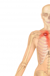
Pain Referral
The Costoclavicular Ligament refers similarly to the Posterior Sternoclavicular Ligament, referring pain deep within the upper thorax. Some Patents have even described the feelings of anxiety and claustrophobia accosted with dysfunction/compression of this structure. Upon effectively treating the Costoclavicular Ligament, they have commented on a reduction of both.

Patients complaining of symptoms similar to and/or test positive for Thoracic Outlet Syndrome, should have their forced coupling mechanism assessed at the SC and CC joints.
I always include treatment of this structure in all of my Patients who present with Anterior Thorax complaints, TOS, impact injuries involving the Shoulder, MVA’s and general shoulder complaints.
Treatment

The costoclavicular ligament is connected to the upper mediastinum, the endothoracic fascia, transversus-thoracicus fascia, parietal and visceral pleura. It also has connections to the mediastinum, fibrous pericardium, phrenic nerve and diaphragm.
Contact this ligament at the sternal attachment of the 1st rib just posterior and inferior to the clavicle.
Make sure you are NOT on the second rib located at the Manubriosternal angle.
Matching the reciprocal tension of the tissues with your flat thumbs, load your pressure posteriorly, inferiorly and laterally while your patient exhales. Repeat and gently hold this position until the tissues soften. Once a softening occurs, reassess for suppleness in the tissues.
HINT
If you have patients with dysfunctional forced coupling of the SC Joints, look to treating this ligament first and reassess. I have found that many times, the dysfunctional SC pattern is due mostly or in part to this structure influencing it.
Earn and Learn!
If you are interested in learning more about how to apply LASTechniques to the shoulder, here’s where you can learn!
Treatment of the Costoclavicular Ligament
Enjoy!
Robert