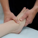
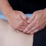
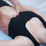
In my 18yrs of practice, I’ve gained the perspective and feel that it is important for the manual therapist to have knowledge of the broad range of injuries that occur to the wrist and leg, to be familiar with appropriate treatment, conservative management, and to refer out when appropriate.
This article will focus on the distinct, immediate and in some cases ongoing injuries of the much less understood Interosseous membranes (IOM).
The interosseous membrane is located both in the forearm and also the lower leg. It is a structure that has ligamentous characteristics. The membrane provides stability for the radius and ulna and for the tibia and fibula. It provides assistance in the maintenance of longitudinal stability and correct functional position. When an injury occurs to the interosseous membrane, a decrease in stability is created in these complexes and a lack of functionality ensues.
Mechanoreceptors of the IOM
Various study’s and reports differ on which types of receptors are present in the interosseous membranes. In cats and rats, the most abundant receptors have been the Pacinian receptors. These are usually located at the periosteal connections of the IOM attachments.
Pacinian corpuscles here seem to be responsible for monitoring vibrations transmitted fro the ground or elicited by jumping or walking. They are also possibly involved in registering vibrations produced by the irregularities of a surface when fingertips are drawn across it and may contribute to the discrimination of different textures.
One paper also reported finding Ruffini-like endings innervating fibrous tissues throughout the body, including interosseous membranes, tendons, tendon sheaths, muscle fasciae, joint capsules and ligaments.
Ruffini-like endings have been shown to determine tensile stress in collagenous bundles. These afferents not only respond to fingertip forces but can also respond to the flexion state of the distal interphalangeal joints, which presumably influences the tension in the receptor-bearing collagenous strands.
The IOM of the Upper Extremity
The IOM of the forearm is composed of three portions:
- a central tendinous tissue
- a membranous tissue
- a dorsal oblique cord
In one study, the distal oblique cord was found in less than half of the subjects. Those with the distal oblique cord had greater stability in the distal radioulnar joint.
The IOM, in particular the distal portion of the IOM, plays an important role in constraining dorsal dislocation of the radius at the Distal Radioulnar Joint.
The IOM is stretched when the wrist is in neutral position, and flexed at pronation and supination positions. Most injuries to the IOM occur along the ulnar insertion. In half of the injuries, there is simultaneous dorsal oblique bundle disruption.
The IOM of the Lower Extremity
Sonography shows the IOM appeared as a thin, hyperechoic (nearly equal to bone cortex) line, continuous from the tibia to the fibula.
It is continuous below with the interosseous ligament of the tibiofibular syndesmosis, and presents numerous perforations for the passage of small vessels.
It is made up of two fibrous networks.
- a major one composed of large diameter fibers grouped into bundles
- and a fine filamentous network
Mechanical studies have shown that the interosseous membrane of the leg is stronger, but less extensible, parallel to the direction of the main fiber bundles, which therefore probably have a load bearing function.
The anterior and posterior inferior tibiofibular ligaments, the interosseous ligament, and the interosseous membrane act to statically stabilize the distal tibiofibular joint. A taut interosseous membrane results when the flexors of the foot contract during stance phase.
Muscular Attachments
The muscles listed below have attachments onto their respective IOMs.
Forearm
flexor digitorum profundus
flexor pollicis longus
The pronator quadratus also helps the interosseous membrane to hold the radius and ulna together, particularly when upward thrusts are transmitted through the wrist.The Abductor Pollicis Longus Muscle
The Extensor Pollicis Brevis Muscle
The Extensor Pollicis Longus Muscle
The Extensor Indicis Muscle
Leg
Extensor Digitorum Longus
Tibialis Anterior
Extensor Hallicis Longus
Peroneus (fiburlaris) tertius
The Flexor Hallucis Longus Muscle
The Tibialis Posterior Muscle
It’s quite obvious, to me at least, that dysfunction tensional differences between the muscles, place tensional stress onto the IOM. This possibly contributes to common complaints of pain/tension/dysfunctions of the wrist and ankle complex’s. I have anecdotal documentation of treatments utilizing L.A.S.T. and Direct Fascial Mobilization techniques to these muscular and ligamentous structures, having a decrease in patient signs and symptoms.
Pathologies/Injuries
Below are a few examples of injuries that can occur to the IOM and related tissues.
When the interosseous membrane is damaged, it may be impossible to detect the problem on any diagnostic test. X-rays and MRI’s are not established to detect subtle changes in the strength of the ligament.
Injuries where the Distal Radioulnar Joint (DRUJ) has been dislocated have been shown to also result in the distal IOM rupture. The IOM, in particular the distal IOM, plays an important role in constraining dorsal dislocation of the radius at the DRUJ. The distal IOM limits palmar and dorsal laxity of the radius at the DRUJ in all forearm rotation positions.
Syndesmosis is a very common injury and is often misdiagnosed. Recurrent or chronic pain in the wrist, elbow, ankle or knee is often associated with damage to the interosseous membrane. The most common mechanisms are a combination of external rotation and hyper dorsiflexion.
Syndesmosis of the Wrist
This type of injury is common for those who perform gymnastic moves or those who have fallen or have had any impact to their lower legs or forearms. Any field, court or ice athlete can obtain this injury from a fall. Any heavy lifting done at too early of an age will result in this type of injury. Tennis Elbow or Pitchers Elbow often is a misdiagnosed syndesmosis.
Figure athletes specifically spend a large amount of their time, doing impact cardio and this can result in a syndesmosis. This type of injury also occurs with bodybuilders or other athletes who engage in lifting heavy weights, especially if the activity continues while they have a growth spurt or underlying injury.
Syndesmosis of the Ankle
The syndesmotic ligaments, responsible for maintaining stability between the distal fibula and tibia, consist of the anterior tibiofibular ligament, the posterior tibiofibular ligament, the transverse tibiofibular ligament, the interosseous ligament, and the interosseous membrane (Figure 10-1b). Injuries to the ankle syndesmosis occur as a result of forced external rotation of the foot or during internal rotation of the tibia on a planted foot.
Anterior Interosseous Nerve Compression Syndrome
The anterior interosseous nerve arises from the median nerve (C8 and T1)
The anterior interosseous nerve (AIN) is only a motor nerve innervating the deep muscles of the forearm. This nerve classically innervates 2.5 muscles:
- flexor pollicis longus
- pronator quadratus
- the radial half of flexor digitorum profundus (the lateral two out of the four tendons).
This nerve can be affected by either a direct penetrating injury or compression. Symptoms involve weakness and possible paralysis of the flexor digitorum profundus, the flexor pollicis longus and the pronator quadratus muscles without sensory loss.
If no signs of spontaneous recovery appear within 12 weeks, operative treatment should be performed.
Posterior Interosseous Nerve (C7 and C8)
This nerve is the continuation of the deep branch of the radial nerve, after it has crossed the supinator muscle.
It supplies all the following muscles on the radial side and dorsal surface of the forearm:
- Extensor carpi radialis brevis – deep branch of radial nerve
- Extensor digitorum
- Extensor digiti minimi
- Extensor carpi ulnaris
- Supinator muscle – deep branch of radial nerve
- Abductor pollicis longus
- Extensor pollicis brevis
- Extensor pollicis longus
- Extensor indicis
The PIN may be entrapped at the Arcade of Frohse, which is part of the Supinator muscle. Posterior interosseous neuropathy is purely a motor syndrome resulting in finger drop, and radial wrist deviation on extension.
Posterior interosseous nerve palsy is a rare syndrome frequently unrecognized, while the clinical presentation is characteristic of finger extension paresis associated with wrist extension in radially deviated position.
Essex-Lopresti Injury
The Essex-Lopresti injury is a rare complex injury of the forearm consisting of a fracture of the head of the radius, rupture of the interosseous membrane and disruption of the distal radioulnar joint.
It is usually the result of a fall on the outstretched hand. A longitudinal force is transmitted through the wrist to the head of the radius which is fractured. If sufficient force is exerted the head of the radius will dislocate, after which the interosseous membrane ruptures, the distal radioulnar joint is disrupted and the radius migrates proximally leaving the patient with a complex instability of the forearm.
The injury is divided into Types I, II, III depending on the severity of injury. More often than not, this injury requires reconstructive surgery. We as manual therapists will possibly see this in our offices post surgery.
Specific treatments for the Interosseous membrane and surrounding tissues are covered in our Upper Body & Extremities course.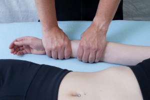
Conclusion
Armed with good clinical knowledge of anatomy and an understanding of common upper and lower extremity injuries, manual therapists can successfully manage many patient complaints.
In my office I regularly refer to our Naturopathic Physician for Prolotherapy treatment in cases of extreme ligament and capsule laxity. Every technique has its place and time and I have found that combining these modalities has only lead to greater improvements in stability, kinesthetic and proprioceptive efficiency and decreased patients complaints. I suggest to you that including a skilled physician who treats utilizing Prolotherapy into your referral network, will only enhance your practice and more importantly, your patients lives.
As always, I hope that I have been able to serve you in some way with this information, be it a completely new perspective into your patients, be it some new thoughts about how to approach a treatment or provide you with a review and acknowledgement that what you are doing, as far as I’m concerned, is having an amazingly positive impact in improving the quality of life for your patients.
Register Now!
References
1. Anatomical and biomechanical study on the interosseous membrane of the cadaveric forearm.
http://www.ncbi.nlm.nih.gov/pubmed/21635800
2. Forearm interosseous membrane trauma: MRI diagnosis criteria and injury patterns
http://www.springerlink.com/content/q5th028086801w06/
3. Learn more about Syndesmosis.
http://www.bodybuilding.com/fun/drryan26.htm
4. Contribution of the Interosseous Membrane to Distal Radioulnar Joint Constraint
http://www.jhandsurg.org/article/S0363-5023(05)00459-4/abstract
5. http://www.ncbi.nlm.nih.gov/pubmed/22245280
6. Sonographic evaluation of lower extremity interosseous membrane injuries: retrospective review in 3 patients.
http://www.ncbi.nlm.nih.gov/pubmed/14682426
7. The mechanical and structural characteristics of the tibio-fibular interosseous membrane.
http://www.ncbi.nlm.nih.gov/pubmed/1274549
8. Dynamic fibular function: a new concept.
http://www.ncbi.nlm.nih.gov/pubmed/954295
9. Muscles of the Leg
http://download.videohelp.com/vitualis/med/mmleg.htm
10. Muscles of the Forearm
http://download.videohelp.com/vitualis/med/mmforarm.htm
11. Slowly Adapting Mechanoreceptors in the Borders of the Human Fingernail Encode Fingertip Forces
http://www.jneurosci.org/content/29/29/9370.full
12. A Consideration of the Elbow as a Tensegrity Structure
http://www.tensegrityinbiology.co.uk/articles/elbow.html
13. Nerves and Mechanoreceptors: The Role of Innervation in the Development and Maintenance of Mammalian Mechanoreceptors

